Porcine Respiratory and Reproductive Syndrome (PRRS)
A page about porcine respiratory and reproductive syndrome (PRRS) describing cause, clinical signs, diagnosis and control.
Information
This disease was first recognised in the USA in the mid 1980’s and the causal agent was identified in the early 90’s in Europe. It was initially called “mystery swine disease” and subsequently was variably named blue ear disease and Porcine Respiratory and Reproductive Syndrome (PRRS).
Aetiology
PRRS is caused by a small, enveloped RNA virus that is classified as a member of the genus Arterivirus of the family Arteriviridae. Genetic, antigenic and virulence differences exist between American and European isolates.
Epidemiology
Transmission is by oro-nasal exposure, via semen or needles. The virus is shed for 1-2 months in young animals (up to 2 weeks in adults) in nasal secretions, saliva, faeces and urine. Transmission is usually by close contact between animals though it can spread via contaminated equipment. Clinically normal pigs can be persistent carriers of the virus. The virus is transmitted vertically in utero or postpartum. Horizontal transmission occurs to naïve animals at later stages of production. Maternally derived antibodies provide some but not total protection. Herd to herd transmission can occur by the introduction of carrier animals or the use of infected semen. However, boars only shed virus in semen for 3-4 days. Sows infected at 85-90 days of pregnancy may give birth to infected piglets. These piglets may shed the virus for a prolonged time. At colder temperatures the virus can last for up to a month. In the environment, infectious virus was rapidly inactivated by drying ; a humid environment is crucial for maintaining virus viability. However, there are reports of spread up to 3 km. Another important issue is the high re-infection rate often without any indication of the source of the new virus.
Clinical Signs
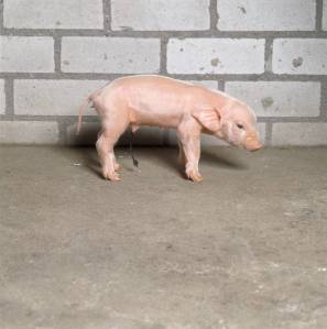
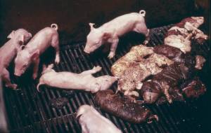
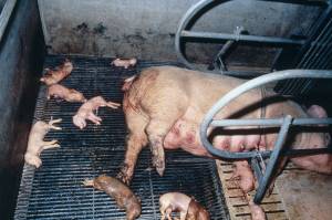
PRRS should be considered when there are clinical signs of respiratory disease at any stage of production, in herds with reproductive problems and in herds with suboptimal performance. Disease is often subclinical and immunosuppression, caused by the virus, can lead to other disease manifestations. Co-infection with Mycoplasma hyopneumoniae results in more severe and prolonged PRRS virus-induced lung lesions. PRRS virus infection has also been shown to increase the morbidity associated with Haemophilus parasuis and Streptococcus suis infections. Clinical signs caused by uncomplicated acute PRRS virus infection may be seen in both sows and piglets and may cause fever and sick animals. Disease may progress through the herd in waves of illness spaced 3-4 months apart with outbreaks only seen in naïve herds. The signs are decline in milk yield / agalactia in sows, increased numbers of late term abortions and weak or stillborn piglets, and increased mortality in the nursing period. Neonatal piglets can show respiratory signs, failure to thrive and increased mortality. Grower pigs often contract pneumonia and are susceptible to secondary bacterial infections like meningitis (Streptococcus suis). The severity of the clinical problems varies greatly among herds and some herds become infected without showing clinical signs. Severity depends on the immune status of the herd, the strain of virus and management factors. Signs are mainly seen in growing pigs where disease has become endemic.
Diagnosis
Clinical signs
Clinical signs of PRRS are distinctive though not diagnostic. Clinical severity can vary from mild to severe and the disease can present as different syndromes attributable to immunosuppression by the field virus. Diagnosis should be confirmed by a variety of tests discussed below.
Post mortem
Serum, lung (tissue or wash fluid) are the best samples to submit to the lab from acute cases while tonsil and lung wash fluid are better for chronically affected animals. Weak newborn piglets are better for virus isolation than mummified or aborted foetuses. To find this virus the Polymerase Chain Reaction (PCR) or virus isolation can be used. The latter is laborious, time consuming and not routinely conducted by many labs.
Virus isolation (VI)
Pigs can be viraemic for 4 weeks but still it is not very easy to sample the right animals. Pigs with clear infection can best be sampled 4-6 weeks after onset. Be aware that MLV-vaccinated piglets can also be positive as the vaccine-virus remains long present in the vaccinated piglet. If needed vaccine virus can be differentiated from field viruses by sequencing the ORF segments. Another practical method to separate field virus from Porcilis PRRS vaccine virus is by the type of cell’s where they grow on. Field virus grows only (>95 %) on Porcine Alveolar Macrophages (PAM cells) where European-strain live PRRS vaccine virus grows only (>95 %) on so called MARC cells. Piglets vaccinated with this vaccine have a positive VI result from 2-6 weeks post vaccination. Adult animals have much shorter viraemic periods and consequently detection and diagnosis can be challenging.
PCR
This technique detects the genetic part of the virus and is therefore very specific. It is an easy technique as compared to VI but it detects any genetic part including that of dead virus. PCR tests are very sensitive detecting also very low quantities of virus.
Both for VI and PCR the demonstration of the virus alone is not very meaningful. Most animals harbour the virus without clinical consequences so finding the virus must be associated with clinical signs to assert the diagnosis. For example when virus is detected in aborted foetuses or fresh weak-born piglets, PRRS is likely to be the cause of the same. Also here we have to be prudent that the PCR can be positive due to the vaccine virus.
Serology
Serology is probably the most widely used diagnostic intervention but has some deficiencies. First of all titres are not directly related to protection. Tests only detect virus neutralizing antibodies. Piglets after vaccination will not all show seroconversion. Antibody levels in sows, that repeatedly came in contact with the field or vaccine virus, will gradually become low in titre or even negative.
So an individual pig that is seronegative can still be persistently infected with virus.
The IFA/IPMA test is the gold standard . Results are expressed in 2 log titres. Every step in titres means a doubling of the titre. The most used test however is the ELISA. The results are expressed as a SP ratio. Cut off level is considered to be 0.4. Every increase by 0.4 is correlated to one titre step in the IPMA test.
After repetitive sow vaccination the expectation is to have low titres where there is no further circulation of field-virus This can be expected 5-6 months after start of vaccination program in the herd. At the start with sow-vaccination there are still booster effects due to the relatively novel heterologous contact.
After piglet vaccination you can expect values similar to those after contact with field-virus.
The ELISA test becomes positive 1-2 weeks after contact or vaccination. Highest titres appear around 5-6 weeks. After this, titres go down again and many animals can become seronegative in time (after some months) The sensitivity of the ELISA is around 95% so the chance of an individual false-negative result is 5%.
After vaccination of piglets with MLV vaccine not all will seroconvert. It is very common that 20-40% do not seroconvert though were protected in challenge studies.
When pigs are infected or vaccinated with homologous PRRS-virus no increase of titre will appear. So after repetitive vaccination (dead or MLV-vaccine) titres will not increase. When titres do increase this is an indication that the sampled pigs had contact with heterologous field-virus.. Dead vaccines elevated titres to approximately 1.5. Higher titres indicate PRRS field-virus contact. MLV vaccines push titres up to around 3.5. Antibody levels from natural exposure can persist for 4-10 months after infection.
Control
The first step in control is to determine the prevalence of infection and timing of virus transmission. This can be done by bleeding groups of pigs for serology to determine patterns of infection. Various management approaches can be taken to the different stages of production as follows:
Nursery management
• Prevent the introduction of infected stock into the nursery
• Euthanasia of weak pigs as these could be persistently infected
• Maintain a strict all-in/ all-out flow in the nursery
• Partial depopulation of weaners coupled with vaccination
• Vaccination of adult stock with a modified live vaccine
Partial depopulation
• Partial depopulation is an effective means for eliminating PRRS virus from endemically infected weaned pig populations in farms with segregated production, once it is implemented in association with a program of elimination of the virus within the breeding herd. It achieves best results when used in conjunction with vaccination.
Whole herd depopulation-repopulation
Whole herd depopulation-repopulation has been widely used in the industry. While it is possible to eliminate PRRS virus using this strategy, maintaining the farm PRRS virus-free
depends on the status of the incoming replacement stock.
Test and removal or Herd closure
Successful elimination of PRRS virus by test and removal has been described in a number of commercial farms. Herd closure is an alternative to test and removal. This protocol is based on preventing the entry of replacements for 4-8 months halting spread of the infection within the endemically infected breeding herd, and removing carriers over time
McREBEL technique
McREBEL stands for Management Changes to Reduce Exposure to Bacteria to Eliminate Losses and was proposed by McCaw in 2000. It consists of a series of steps to reduce the spread of PRRS virus among piglets most frequently in herds undergoing acute outbreaks. The advantages of the protocol are its simplicity and low cost. However it was designed in a time when useful modified live vaccines were not available and vaccination has now become centre stage in PRRS control.
Vaccination
Animals may be vaccinated in young life to prevent or reduce the risk of infection with field virus. Additionally now breeding animals that have been shown as already exposed to PRRS virus can be vaccinated with modified live vaccine. A variety of vaccines are available. Your local veterinary practitioner is best placed to advise you in this regard. Further information on the vaccine from MSD AH may be found by clicking here for Republic of Ireland and for Northern Ireland please click here.