Bovine Respiratory Disease
Introduction
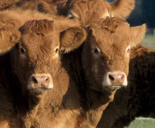
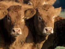
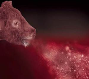
Bovine pneumonia or Bovine Respiratory Disease is a devastating condition affecting cattle of all ages. It is the most common cause of mortality in all ages of cattle except young calves. It is most often associated with the assembly of large groups of cattle from diverse origins. The disease is of considerable economic significance in feedlots, it is responsible for a high mortality rate and the condemnation of infected carcasses at slaughter.
Many factors including stress and management play a role in bovine pneumonia. The most important infectious agents involved are viruses and bacteria, though parasites occasionally contribute to clinical disease.
The aetiology (cause) of bovine pneumonia
This disease is multi-factorial including the following causes:
1. Stress – is a major predisposing factor to Pneumonia outbreaks. Stress can arise from:
Passage through marts
Commingling
Weaning, castration, dehorning
2. Infectious agents
Mannheimia haemolytica and Pasteurella multocida are frequently isolated from the lungs of sick cattle. These pathogens are associated with a high mortality rate.
Other pathogens that may also be responsible for disease including:
Respiratory viruses:- RSV, PI3, BoHV-1 (IBR), BVD, Bovine coronavirus
Other bacteria e.g. Histophilus somni, Mycoplasma spp.
Parasites e.g. lungworm may also contribute to disease. For further information on lungworm infection please click here.
Our UK colleagues have also produced a dedicated website with further information: www.huskvac.co.uk.
3. Management
The environment in which animals are kept, nutrition and management of animals may all contribute to increases in pneumonia incidence on individual farms.
Epidemiology (Factors influencing occurrence of disease)
Respiratory disease incidence is dependent on three major factors:
Animal factors
Colostrum is depleted by 3 months of age so passive protection waning leads to higher prevalence in this age group
The efficiency of an animal’s immune system may be reduced by stress from certain farming activities such as weaning, dehorning, castration, transport or mixing and change in dominance hierarchy.
The age also influences incidence as the immune system and lungs are not fully developed until 12 months old.
Environmental factors
Air quality – Irritant dust & gases depress respiratory defences
Draughts stress animals
Humidity – High humidity favours dampness & disease spread.
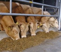
Population density – Infective potential of respiratory disease increased with crowding.
Mixing of age groups facilitates disease spread from carrier animals
Mixing of animals with different disease exposure history facilitates spread to animals not previously exposed to that agent in the group.
Infectious agents – viruses, bacteria and parasites
Direct damage can be caused by certain infectious organisms such as BRSV, Mannheimia haemolytica, IBR virus, Pasteurella multocida and Histophilus somni. Parasites (lungworm) can also contribute to significant disease and can lead to secondary infections with bacteria etc.
Other agents predispose to damage such as PI3 virus, BVD virus, Mycoplasma dispar, Mycoplasma bovis and other viruses.
Clinical Signs
During an outbreak of bovine pneumonia, mortality in a herd can be as high as 25%. The disease is responsible for up to 75% of morbidity in groups of housed cattle and is the primary threat to newly weaned and newly brought-in calves. Death and treatment costs can easily attain 8% of production costs. To this must be added the costs of reduced feedstock performance, sub-clinical illness and higher personnel costs.
Clinical signs are influenced by the age of the animal, the agent concerned and whether other agents are also present.
Signs relating to Respiratory Disease
Fever (as high as 42 C)
Depression, laboured breathing and increased respiratory rate
Loss of appetite and cough
Reddening of the mucous membranes
Nasal discharge – Initially watery and later may become purulent
Conjunctivitis – runny eyes
Drop in milk production

Diagnosis
Diagnostics in respiratory disease can be based on
(i) clinical examination
(ii) laboratory testing
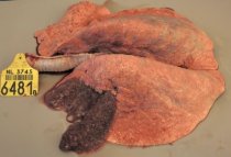
(iii) post mortem examination
(iv) preferably a combination of these different disciplines.
The clinical examination of diseased animals includes
· recording of respiratory rate and type
· rectal temperature
· observation for clinical signs
Clinical signs and gross pathology in respiratory disease rarely yield a specific diagnosis. Moreover, mixed infections are the rule rather than the exception. Laboratory testing is often required for a proper diagnosis.
Laboratory tests can be carried out on samples collected
- from live diseased animals
- during post-mortem examination
Most veterinary diagnostic laboratories offer methods for antibody testing for the common respiratory pathogens. Antibody testing for the different respiratory pathogens can be done by different methods like virus neutralization tests or antibody ELISA. The testing should be done on paired blood samples, because detection of antibodies in a single sample has little diagnostic value.
Samples may also be taken from diseased animals using the following specialist methods:
Nasal swabbing
LUNG WASHING: Two types of lung washing may be carried out: Trans Tracheal Lavage (TTL), involving insertion of a tube through the skin into the trachea or Bronchio-Alveolar Lavage (BAL), where the tube is inserted into the trachea through the external nares (nose). Your vet is best placed to conduct either of these diagnostic procedures.
Samples should be cooled during transport to the laboratory and transport times should be as short as possible
Lungworm assessment of faeces while useful in young grazing animals, may not be entirely accurate in detecting patent infections in adult stock. For further information on diagnosis of lungworm infection click here.
Control
Control of Bovine Respiratory Disease is based on four equally important aspects:
Vaccination – The use of vaccines confers clinical protection and more importantly reduction of the pathogen circulation. Due to the complex cause of the disease, multivalent vaccines are preferred.
A number of live and inactivated vaccines with different combinations of viral antigens are commercially available. Bovilis INtranasal RSP Live is available for intranasal administration from the day of birth to calves to protect against BRSV and PI3 viruses. Onset of immunity is 7 days and it provides 12 weeks protection. Bovipast RSP is a vaccine that protects against Mannheimia haemolytica as well as BRSV and PI3 that contains IRP (Iron regulated protein) antigens. These antigens combined with a special adjuvant allow early protection against Mannheimia haemolytica as well as RSV and PI3. Protection is afforded against serotype A1 as well as A6 of Mannheimia haemolytica. Vaccination programs should start at an early age e.g. from two weeks onwards to protect the young calves. In situations, where protection is required for more than one winter season, it is advisable to re-vaccinate the animals at least 15 days prior to the risk period. For older calves that were not vaccinated earlier, two doses should be administered four weeks apart with the course completed at least two weeks prior to weaning. This vaccine is recommended for use at the same time as Bovilis IBR Marker Live. For further information on the control of IBR, please click here.
Vaccines should only be administered as part of an overall control programme. Advice should be sought from your local veterinary practitioner.
Bovilis Bovipast RSP is an inactivated multivalent vaccine for the protection of all classes of cattle, including pregnant and/or lactating animals and young calves against:
- Parainfluenza 3 virus, to reduce infection
- Bovine Respiratory Syncytial virus, to reduce infection and clinical signs
- Mannheimia haemolytica serotypes A1 and A6, to reduce infection, mortality, clinical signs, lung lesions and bacterial invasion of the lung.
Bovipast RSP is licensed for use at the same time as Bovilis IBR Marker Live.
For information on the control of lungworm please click here or go to www.huskvac.co.uk
- Management– Reduction of circulating viruses and bacterial shedding can be achieved with careful attention to detail. Significant issues in need of address include ventilation in housing, drainage, mixing of age groups, overcrowding and parasite control. For further information on ventilation and calf housing and how they can contribute to BRD control please click here.
- Isolation of clinically affected cases and treating with an appropriate antibiotic can also limit losses.
- Biosecurity – Maintaining biosecurity involves avoiding introduction of infected animals into the herd and/or implementing stict isolation / quarantine of introductions until proven negative, and restricting access of livestock to external sources of infection e.g. double fencing is in place at all perimeters, considering carefully sources of biological materials such as embryos, semen etc.
- Treatment – Therapy is limited to the use of antimicrobials and anti-inflammatory drugs. Antibiotics are only effective against bacterial infections. It is often required to start the therapy before results of the bacteriological investigation are available and the resistance patterns are determined. Consequently, the bacteria exhibit increasing resistance to a large number of antimicrobial agents. Preferably, antimicrobials which are active against relevant bacteria should be used.