
A page about worm infestations in dogs describing causes, clinical signs, diagnosis and control.
Introduction
Worms are a well-recognized worldwide issue. They affect pets, wild animals, livestock and even people. Many worms that we are concerned about are internal parasites of which intestinal worms are most important. Roundworms, tapeworms, hookworms and whipworms are the most commonly encountered types of intestinal worms, while other non-intestinal worms can also cause disease. These include lungworm and heartworm, the latter of which are not considered to be endemic in Ireland.
All worms pose a potential lifetime risk to all dogs and some of these worms can also pose a risk to people as well.
We will now discuss the most common intestinal worms that our pets may suffer from in Ireland – Roundworms, Hookworms and Tapeworms.
Roundworms Toxocara spp
Aetiology
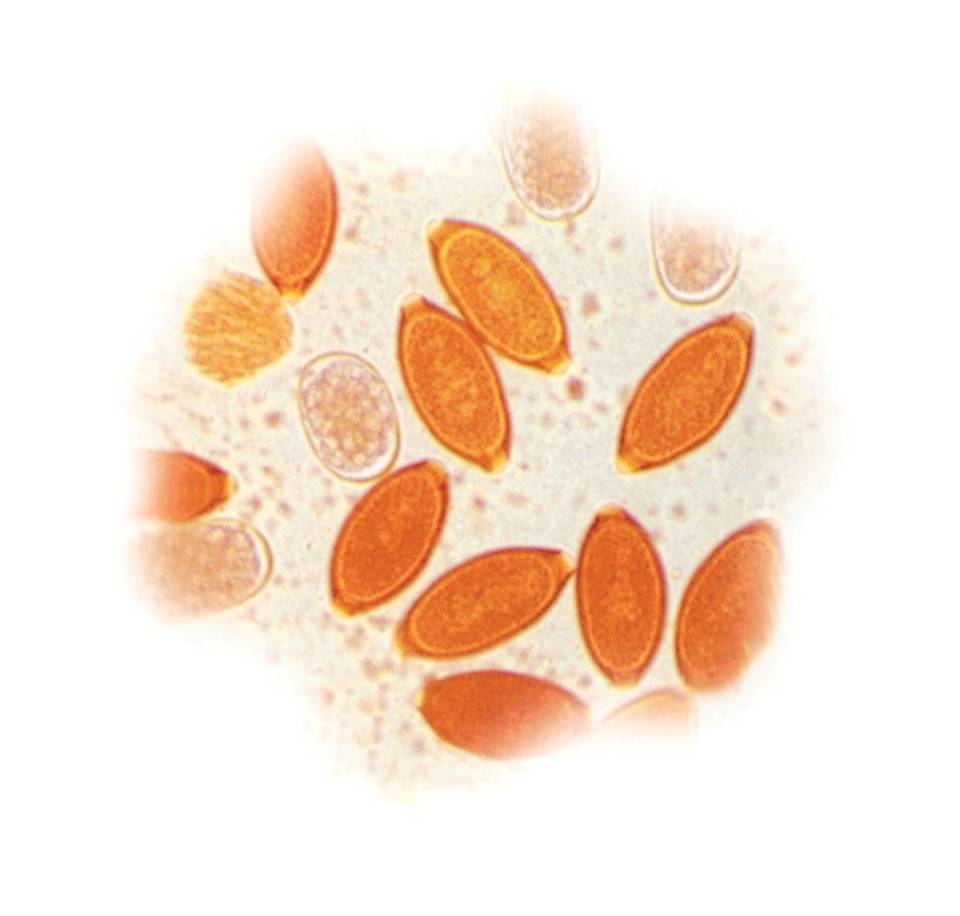
There are over 15,000 known species of nematodes (roundworms), many which simply live in soil. There are two main species of intestinal roundworm which commonly occur in dogs in Ireland and can cause illness in pets and their owners – Toxocara canis and the much more uncommon Toxascaris leonina.
Toxocara canis is the roundworm species which affects dogs. In a recent study of stray dogs in Ireland 6% of the dogs were shedding Toxocara canis eggs!
There are three general stages in the life cycles- eggs or oocysts, larvae and adults. The life cycle is detailed below.
The adult worms live in the small intestine of dogs where they lay eggs, and these are then passed in faeces. These eggs can remain in the environment for months or more and act as a source of infection for dogs. Dogs can also become infected by eating undercooked meat or by eating infected small mammals. These small mammals are known as paratenic hosts, meaning that the eggs can survive in these hosts, without developing into further stages. There are also unique methods of transmission in pups. In pups, infection can occur when larvae cross the placenta or through their mother’s milk.
The eggs hatch in the intestine and develop into larvae. These larvae migrate from the intestines through the liver and lungs and the life cycle is completed when these larvae are coughed and swallowed and then mature into adults in the intestines.
Clinical Signs
In the case of a low burden or in adult animals these infestations are often asymptomatic (show no signs of infestation). However, where infestations become heavy in pups, they may cause severe clinical signs. The clinical signs often include weight loss and a potbellied appearance. More severe clinical implications include the risk of intestinal blockage, intussusception and pneumonia.
Zoonosis
Both species of roundworm pose a significant risk to human health. People can become infected when they come in contact with infective eggs in the environment. As infective eggs are passed in faeces, sandpits and gardens are often sources of contamination. Children and people with compromised immune systems are most at risk. After a person ingests roundworm eggs, the larvae begin to migrate through different tissues. This migration is called Larva Migrans Complex. The larvae can infect different organs such as lungs, liver, brain and eyes. The clinical signs seen can be severe such as blindness or blurred vision and seizures.
Diagnosis and control
Although clinical suspicion might be raised based on the clinical signs seen in dogs, definitive diagnosis is based on faecal analysis.
However, given the risks that these roundworms pose to our pets and family, and the fact that infestations often go unnoticed, it is important to follow expert treatment guidelines. It is recommended that all pups are treated every two weeks from two weeks of age until two weeks after weaning and then monthly to six months old.
Nursing bitches should be treated concurrently with their offspring.
All adult dogs should be treated for worms at least four times a year.
However, in households with young children or immune-compromised people, then this should be done every four weeks.
Hookworms Ancylostoma spp. and Uncinaria spp
Hookworms are small intestinal worms. They are named as they have large mouth parts which are at an angle to the rest of body. There are several different species of note in Ireland. Ancylostoma caninum and Uncinaria stenocephala are the main hookworms in dogs.
However, hookworm infestations are more common in dogs in Ireland. During a recent study 10% of stray dogs in Ireland were shown to be shedding hookworm eggs, making hookworm one of the more common worm which infest dogs in Ireland.
Adult worms inhabit the small intestine with eggs passed in faeces. These eggs develop into larvae which are either ingested or penetrate through the skin to infect new hosts.
Ancylostoma spp species can also be transmitted via the dam’s milk to her offspring.
Clinical signs
Hookworms feed by grasping the mucosa of the intestines and can damage the surface. This feeding action results in the most common signs seen which include diarrhoea, which often contains blood, weight loss and anaemia. Animals infected via skin penetration may also show skin lesions.
Immunity can develop after exposure, although it is unlikely to be full immunity.
Zoonosis
It is considered very low risk as cases have not been reported.
Diagnosis and control
Diagnosis is made by identifying hookworm eggs in faeces. However, in young pups, diagnosis can be complicated by the fact that clinical signs may become apparent before the infection is patent i.e. before the infection is mature and producing eggs which can be found in the faeces.
If a diagnosis of hookworm is made, that animal should be treated with an appropriate anthelminthic treatment and often these patients may need supportive treatment as well.
Control is based on regular worming treatment. There are no specific expert recommendations for hookworm – they are generally considered to be adequately controlled by a comprehensive programme for roundworm control.
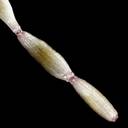
Whipworm
Whipworms are named due to their whip-like appearance – made up of a narrow front end (digestive tract) and thick posterior (containing the reproductive organs). One species affects dogs in Ireland – Trichuris vulpis which is substantially smaller than the other worms (a mere 2-5 cm in length) and is rarely seen as it lives in the caecum (the portion of the large intestine where the small and large intestine meet). In heavy infestations it extends into other areas of the large intestine. The risk to humans is somewhat equivocal. There are anecdotal reports of zoonotic infections though it is thought that the worm does not go through full development in human hosts.
Similar to roundworms, adult whipworms produce environmentally resistant eggs, which are extremely difficult to destroy. These eggs are laid in a non-infectious form and develop to the infective first-stage larvae (L1), still contained inside the egg, in approximately one month. After a dog ingests eggs containing infective first-stage larvae, they hatch and will develop into reproductively mature adults after approximately 3 months. This time, termed the pre-patent period, is remarkably long among the worm species which affect dogs. Unlike roundworms however, whipworms do not migrate outside of the intestinal tract i.e. there is no somatic migration into tissues, no migration of larvae to puppies in utero, and no transmammary transmission to pups from their dam via suckling. Because of the long prepatent period, whipworms are normally diagnosed in dogs older than 6 months.
Clinical signs
Small numbers or a light infection of whipworms generally do not cause clinical signs for the host but if large numbers of worms embed their anterior ends in the large intestinal mucosa, significant inflammation, caused by the irritation of the gut lining during feeding, can lead to a bloody and mucoid diarrhoea. This commonly becomes chronic and hard to control though blood loss is rarely significant enough to cause an acute anaemic crisis. A second syndrome of infection has emerged where dogs show signs similar to Addison’s disease (hypoadrenocorticism). Here, a syndrome of episodic weakness with inability to conserve salt ultimately can lead to a serious dehydration outcome.
Diagnosis
Diagnosis may be suspected based on clinical signs and history. Laboratory methods include flotation of eggs (which are only visible under a microscope but have a unique appearance – see image below) which can be difficult. This is because eggs are shed intermittently or in low numbers and the eggs do not float well. A centrifugal float using Sheather’s sugar solution (specific gravity 1.25 to 1.30) provides the best chance for diagnosis though not all laboratories may conduct this type of flotation and it may have to be requested. In recent years, tests have become available that detect T. vulpis antigen in faeces even before egg shedding has begun. These tests are especially useful for animals with clinical signs but no eggs apparent on faecal flotation.
Treatment
Treatment with anthelmintics is generally effective. Because of the 3-month prepatent period, during which immature whipworms are not susceptible to anthelmintics, treatment administration should span at least three months. It is important to check that the anthelmintic chosen is effective against whipworms. Certain products marketed as effective against internal and external parasites are not effective against whipworms.
Tapeworm
There are a few different tapeworm species that affect dogs in Ireland. Overall prevalence of tapeworm infestation is thought to be less than 7%. There are two species of tapeworm that will be discussed below.
Dipylidium caninum– which is known as the flea tapeworm and Taenia spp.
Dipylidium caninum – Flea tapeworm
This tapeworm can infect both dogs and cats. Fleas and lice are intermediate hosts to the larval stages. Proglottids or tapeworm segments are passed in faeces. These contain oncospheres which are ingested by flea larvae and lice. Dogs are infected when they ingest adult fleas or lice while grooming. The adult tapeworm develops in the intestines of dogs after ingestion.
Clinical signs
Infestations are often totally asymptomatic. However anal pruritus and proglottid segments may be seen in faeces or in the coat around the anus.
Diagnosis and control
Diagnosis can be suspected based on clinical evidence of tapeworm infestation, but again definitive diagnosis is based on faecal analysis.
The control and management of Dipylidium caninum is centred on flea control. Products that encourage compliance regarding flea cover, by providing longer periods of activity, are more likely to produce the best year-round control.
If infestation is suspected or confirmed, then treatment with an appropriate worming product is required.
Taenia spp
This tapeworm can infect dogs, cats and foxes. They are infected by ingesting infected intermediate hosts or by eating infected meat. These hosts are themselves infected by ingesting infective eggs within the proglottids. Different Taenia spp infect different intermediate hosts. Dogs who eat undercooked meat or scavenge animal carcasses are at risk of becoming infected with the following: Taenia multiceps or Taenia hydatigena.
Clinical signs
Like infestations of Dipylidium caninum, infestations can often be asymptomatic. Occasionally signs of anal pruritus might be evident or proglottids may be seen in faeces or on the coat around the anus.
Diagnosis and control
The diagnosis can be suspected given an animal’s lifestyle and habitats, such as hunting or scavenging. A definitive diagnosis is based on faecal analysis. However, faecal analysis has its limitations when identifying tapeworm species as Taenia spp cannot be distinguished from Echinococcus spp. Luckily Echinococcus spp are not present in Ireland.
The control of Taenia spp infestations is based around prevention of infestation and treatment of existing infestations. At-risk dogs should therefore be treated at appropriate intervals.
Prudent use of anthelmintics should be practised in accordance with expert guidelines. This is especially important for products containing praziquantel as this is the only product licensed for all of the common tapeworms. Over-use or inappropriate use can encourage the earlier development of resistance to wormers. Therefore, animals should be treated on a risk basis. It is important to discuss risk factors with your vet and decide on the most appropriate treatment schedule and product choice based on your pet’s risk profile regarding worms.
Lungworm infections
Lungworm infections in dogs are normally attributed to infection with the parasite of the lung vascular system, Angiostrongylus vasorum. However, this a parasite of the vasculature (See later) while the more classical airway associated worm is the tracheal worm Oslerus osleri. This worm occurs in Europe and rarely in Ireland. Adult lungworms live in nodules in the trachea of dogs, and larvated eggs laid by adults hatch there. Pups become infected from the faeces or saliva of an infected dog. Clinical signs range from asymptomatic through moderate, dry coughing with slightly increased respiratory rates to severe, to persistent coughing and respiratory distress. Diagnosis of lungworm infection is based on signs and presence of larvae in faeces (though samples may often be negative in infected animals). Examination of the airways with an endoscope (bronchoscopy) and x-rays can be helpful tools. Bronchoscopy can be used to collect washings from the trachea to examine for eggs, larvae, and white blood cells.
Treatment
Lungworm infection in dogs can be difficult to treat, but there is evidence that appropriate antiparasitic drugs are effective, when combined with surgical removal of the nodules in the trachea. It may be necessary to continue antiparasitic treatment for up to 2 months to effect a cure however.
Angiostrongylus vasorum
Life cycle
The adult form of this worm resides in the vasculture, most commonly of the pulmonary system. It has a somewhat complex lifecycle which begins when the third stage larvae are ingested by a definitive host, primarily the fox or dog. This can be through eating mollusc (intermediate hosts), frogs, lizards, mice and rats (paratenic hosts), or from food infected with slime from the slugs or snails. These larvae (L3) migrate to the lymph nodes draining the bowel and moult to L4, and L5. The L5 larvae migrate through the circulation to the liver via the portal vein, then through the liver and the adults end up at the pulmonary artery or right side of the heart (Right ventricle).
The adults then mate and produce eggs. The eggs move to the alveolar capillaries via the circulation and hatch to L1 larvae. The L1 larvae burrow though the alveolar and are then coughed up and swallowed. L1 larvae are therefore passed in the faeces of infected canids. The L1 larvae infect intermediate hosts (primarily slugs and snails) by penetrating the foot of the slug or snail and develop to L3 inside. Adult worms can live for 2 years. The pre-patent period (time between infection and when egg shedding begins) is 6–10 weeks.
Clinical signs
Cardio-respiratory signs can arise as vague clinical signs. Chronic coughing, exercise intolerance, difficulty breathing or fast breathing in young dogs due to blood vessels being blocked by adults, eggs and larvae, can occur. The parasite also causes clotting difficulties. Haematomas and prolonged bleeding are as a result of thrombocytopaenia (a decrease in the number of platelets in the blood) because the parasite produces anti-clotting factors, to ensure its ongoing survival in the vasculature.
Angiostrongylus vasorum also causes nervous signs including ataxia, paresis, loss of vision, behavioural changes and seizures resulting from haemorrhages in the nervous system.
Diagnosis
Diagnosis is made from a combination of clinical signs and tests including chest X-rays, poor clotting ability and a finding of anaemia. Some cases may also have hypercalcaemia of unknown aetiology. Faecal examination using the Baermann technique are unreliable, as egg output is irregular and the pre-patent period of the parasite is relatively long, so disease can be advanced by the time eggs show up in faeces. Multiple samples help reduce the risk of a false negative but should not be relied upon.
Post mortem examination finding the worms and associated haematomas and enlarged blood vessels are diagnostic. Occasionally endocarditis of the right side of the heart and the tricuspid valve are present.
Treatment and prevention
In Ireland a variety of products are approved for prevention of Angiostrongylus vasorum in dogs and certain products are also indicated for treatment of the infection. In endemic areas, monthly application of effective anthelmintcs will prevent angiostrongylosis and patent infection. Your vet will advise you on the necessity for control or not depending on your dog’s risk profile.
Heartworm
Heartworm is mostly prevalent in southern France, Spain, Italy and the Mediterranean. Mosquitoes are responsible for transmitting the disease to dogs and many different species of mosquitoes can carry heartworm larvae. This parasite does not occur endemically in Ireland but occasional cases may arise after return to Ireland that are due to infection occurring while travelling abroad.
Who is at risk?
Irish dogs could be more vulnerable as they have never encountered the disease and therefore have no resistance.
How is it spread?
The larvae of this worm (which eventually resides in the heart) are present in the bloodstream and can be transferred to an unaffected dog via a simple mosquito bite.
The life cycle of heartworm is developed in the following stages:
• Adult heartworms reside in the right heart chamber.
• Heartworm larvae are released into the dog’s blood and the mosquito ingests the larvae with the dog’s blood.
• After 10 to 30 days, the infective larvae appear in the salivary gland of the mosquito, so when the mosquito bites another dog, it transmits the infective larvae.
• The larvae then migrate around the dog’s body for about four months before reaching the dog’s heart. The larvae mature into adult worms over the next three months.
• The process repeats itself.
Signs and symptoms
Heartworm disease is caused by damage from the adult worms once they get into the blood vessels of a dog’s lungs. The worms cause the blood vessels to swell and become scarred. As the blood vessels shrink in diameter, blood flow becomes restricted and blood pressure begins to rise. Eventually, the increasing blood pressure will lead to heart failure.
Signs may take several years to manifest and include soft cough, tiredness, weakness, loss of weight and condition. Eventually heart failure may ensue.
Prevention and control
There are products available in Ireland that may be obtained from your vet prior to your trip, or from a local vet on arrival. They kill the larvae after infection.