Uterine Infection
A page about uterine (womb) infections (post-calving) in cows.
Introduction
Bacterial uterine infections disrupt not only the function of the uterus, but also the ovary and the control centres in the hypothalamus and pituitary gland. The correct diagnosis and treatment of uterine diseases is a key component of all reproduction management programs.
Traditional classification of uterine infections differentiates between acute endometritis (vaginal discharge, enlarged uterus and clinical disease) occurring until 10 days post partum and subacute-chronic endometritis (limited vaginal discharge, absence of clinical signs) occurring within 21 days or more post partum not accompanied by systemic signs. Pyometra is an accumulation of purulent or mucopurulent within the uterine lumen and distension of the uterus in the presence of an active corpus luteum.
Aetiology
Up to 90% of recently calved cows have uterine infections 1 to 2 week after parturition. It is the ability of endometrial defences to eliminate the infection that decides on the progression of uterine infections.
Cows with retained placenta and labour complications show increased bacterial contamination of the uterus. There is a marked difference in the population of bacteria responsible for uterine infections in different periods post calving (See table below). It has been well established now that there is certain correlation between bacterial species involved in uterine infections.
| Bacteria | Acute | Subacute |
| T. pyogenes | 33-83% | 33-85% |
| Gram neg bacteria | 49-67% | 17-70% |
| E. coli | 67-85% | 0-17% |
| Peptostreptococci | 60-80% | <5% |
| Remaining | 23-52% | 17-39% |
Epidemiology
Factors predisposing to uterine infections in cows include risk factors for the establishment of uterine bacterial disease. These have been published by Sheldon and Dobson (2004). They include:
Uterine damage
• Stillbirth, twins, dystocia, Caesarean section
• Retained placenta
• Delayed uterine involution
Metabolic conditions
• Milk fever
• Ketosis
• Left displaced abomasum
Balance between pathogenicity (disease causing ability) and immunity
• Disruption of neutrophil function
• Type of bacterial flora in the uterine lumen
• Progesterone or glucocorticoid (medicine) administration leading to early formation of corpus luteum
• Level of hygiene of the environment – cows or calving boxes may be less important
Clinical Signs
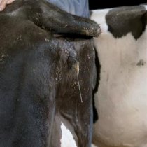
Acute puerperium metritis
A life threatening condition that can become chronic later in the post partum period.
Endometritis
Signs include: morphological and functional changes in the endometrium lead to decreased fertility (delayed submission for AI, repeat breeding). Uterine discharge may or may not be visible externally.
• Disrupted luteolytic path exposing the uterus to the immunosupppressive action of progesterone.
• Decreased milk production.
• Impairment of ovarian function: delay in first ovulation and impaired growth of dominant follicle in cycling cows
Diagnosis
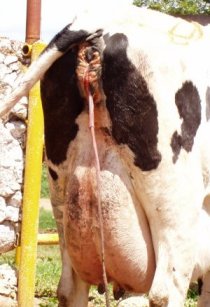
Diagnosis of metritis within the first 10 days post partum is relatively easy. It is associated with the presence of pyrexia, fetid pus within the uterine lumen, vagina and discharging from the vulva accompanied with delayed uterine involution. Clinical and subacute endometritis may be more difficult to recognize. A complete clinical examination sometimes followed by laboratory tests is required for a definitive diagnosis. Clinical examination with detailed evaluation of the reproductive tract
• Rectal examination
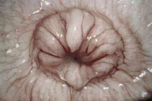
• Vaginoscopy:
Requires additional equipment and provides valuable information: presence of discharge from the cervical canal, condition of the vaginal mucosa, status of the external cervical orifice
The use of vaginsocopy or manual examination of the vagina and mucous discharging from the cervix is thus highly recommended. Manual examination is quick and inexpensive. Additional information such as the presence of vaginal lacerations and odour of the discharge is also obtained. Metricheck (Metricheck, Simcro, New Zealand), a device consisting of a stainless steel rod with a rubber hemisphere, can also be used to retrieve vaginal contents.
Your vet is best placed to carry out all of these procedures.

Additional laboratory investigations are also possible as follows:
• Cytology- of uterine contents gives valuable information and allows for a diagnosis of subclinical cases
• Bacterial culture and sensitivity- Provides important information about the infection and indicates which antibiotic to be used.
Important points:
o Proper sample collection is essential to avoid false positive results (double sheeted swabbing pipette, avoid side contamination from vagina, vulva and environment)
o Transportation of samples
o Bacterial culture (both aerobic and anaerobic) o Interpretation of results
• Biopsy and histology- Provides information on the extent of inflammation, and the health and functional status of the endometrium. Biopsies are helpful for formulating a prognosis.
Important points:
o Sample collection
o Interpretation of results
o Equipment and costs
Your vet is best placed to advise you concerning these procedures.

Treatment
Treatment of bovine endometritis aims at the:
Elimination of bacterial infection
Antibiotics used in the treatment of uterine infections have to comply with certain criteria including high activity against the main pathogens, where injectable formulations must reach high concentrations in uterine lumen and endometrium while intrauterine must result in good penetration in the lumen and endometrium, no irritation to endometrium, no negative effect on uterine immune cells and finally a short milk withdrawal period. MSD Animal Health recommends a specific intra-uterine antibiotic for the treatment of clinical and subclinical endometritis. This product may only be prescribed by your veterinary practitioner from whom advice must be sought.
Improvement of uterine contractility
This results in evacuation of pathological contents of the uterus
Elimination of immunosuppresive influence of progesterone
Role of prostaglandins in the therapy of uterine infections (Lewis 2004):
1. Elimination of corpus luteum which results in:
– improvement of uterine contractility
– elimination of immunosuppressive effect of progesterone
2. Direct stimulation of the function of immune cells in endometrium
Preventing recurrent infections during the following luteal phase
If prostaglandins alone are used in the treatment of endometritis, clinical improvement can be achieved, some bacteria may however remain in the endometrial crypts. These bacteria will multiply during the next luteal phase when the immune function of the endometrium is decreased due to the effect of progesterone. This leads to relapses of endometritis.
Combination of proper anti-infective therapy and prostaglandins allows not only elimination of existing bacterial infection but also prevents recurrence of the condition in the following cycle. Furthermore, Le Blanc found that the use of prostaglandins prior to first ovulation after calving (e.g. pre day 25) may result in a depression in subsequent fertility.