Liver Fluke
A page about liver fluke in cattle, it cause, clinical signs, diagnosis, treatment and control.
Introduction
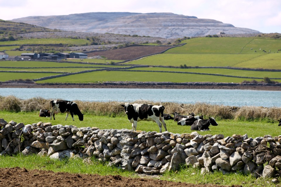
Liver fluke, known as Fasciola hepatica, is a flattened ovoid shaped parasite that, as its name suggests, colonises the liver of various species most notably ruminants. The parasite can also affect other species including horses and even humans. Various stages of disease cause different clinical signs and are often associated with specific seasons of the year e.g. sheep often show clinical signs of acute disease whereas this syndrome is relatively rare in cattle, while signs related to chronic disease are relatively common. Grazing animals are infested through the ingestion of infected grass and the disease is estimated to cost the Irish cattle industry up to €90M per year in production losses.
Aetiology
Liver fluke disease is caused by a trematode parasite called Fasciola hepatica. Various different species of Fasciola occur around the world but only one occurs in Ireland.
Epidemiology
Liver fluke has a somewhat complex life cycle in which adult fluke, present in the bile duct of infected animals lay eggs. These eggs pass out in faeces and under correct conditions of humidity and climatic temperature (>10C) they hatch and the resultant life cycle stage (miracidia) enter a snail. Here they undergo massive multiplication to produce thousands of redia and subsequently metacercariae. These then exit the snail and swim to a convenient part of the grass for animals to eat. Here they form a cyst around themselves for protection against adverse environmental conditions. Grazing animals ingest the encysted metacercariae, which uncoat in the stomach and penetrate the intestine travel via the blood system to the liver. Here they travel through over a period of approximately 10 weeks to reach the bile ducts as adults where they begin the cycle once more. Seasonally the grass becomes infested in the spring / early summer when infected animals are released to grass. Newly grazing animals take in infective metacercariae and this begins the life cycle. As a result, earliest disease (acute disease most often seen in sheep) occurs in August / September. Disease sydromes associated with subacute or chronic infestations occur from November through January. There are exceptions to this life cycle e.g. animals infested for the first time late in the year will obviously not develop chronic fluke until the springtime etc.
The level of fluke infection on a pasture varies from season to season and is dependent on a number of factors:
- Weather in the previous year – from August to October. Mild temperatures and above average rainfall provide optimum conditions for fluke development
- Weather in the current year – from May to July. Above average rainfall provides optimum conditions for fluke development
- Incidence of liver fluke in the previous year. When incidence has been high previously it facilitates fluke development in subsequent years as it contaminates the pasture. Damp muddy areas are more prone to liver fluke infestation as intermediate stages of the life cycle require the mud snail (Lymnaea truncatula) for multiplication
- The choice and timing of flukicides in a control programme – this is discussed further under control
Liver fluke are thought to overwinter through a variety of mechanisms including as adult fluke in animals or as encysted metacercariae in the soil.
Clinical Signs
Clinical signs in cattle may often be more subtle than in sheep. They classically include the following:
- Reduced liveweight gains through reduced feed conversion efficiency
- Reduced milk yields
- Reduced fertility
- Anaemia manifested as pallor of the mucous membranes (linings of the eyes and mouth)
- Bottle jaw – a swelling of fluid underneath the jaw – may clinically resemble “Timber Tongue” though the tongue is not affected
- Diarrhoea
- Severe cases can lead to death. Furthermore the disease can be complicated by Black Disease – a clostridial disease caused by Clostridium novyi.
Often the disease can be detected in animals in the abattoir due to condemnation of livers as a result of damage caused by the fluke travelling through the liver, leaving fibrous tracts throughout the liver substance.

Diagnosis
Clinical signs of liver fluke infestation are suggestive of the disease but laboratory tests are required for a definitive diagnosis. Often animals are not yet shedding eggs or only shedding intermittently and this can lead to a false negative diagnosis. In summary do not use a negative faecal egg count as a reason to rule out the possibility of fluke infestation.
Blood sampling for antibody levels / Bulk milk tank testing
Retrospective diagnosis of liver fluke infection can be made by measuring antibody levels in sera samples. Consult your local veterinary practitioner for further details. A bulk milk antibody test is also available for indication of the presence of infection at herd level.
Control
Control of liver fluke is based on four equally important aspects:
- Improve drainage and fence off muddy areas during risk periods
- Monitor fluke infestation using faecal egg counts, bulk milk tank antibody levels and feedback from abattoir liver post mortem examination – care should be exercised in interpretation of results however. Negative egg counts may not mean freedom from infection.
- Biosecurity – Maintaining biosecurity involves avoiding introduction of infected animals into the herd and/or implementing stict isolation / quarantine of introductions until four weeks have elapsed after treatment with a flukicide effective against immature stages.
- Strategic dosing:
- A strategic early summer dose with Zanil will remove adult fluke and reduce the risk of pasture contamination
- Subsequent advice on summer dosing should be advised based on results of the monitoring programme. This in turn will depend on weather, farm risk etc.
- Once animals are housed a flukicide, effective against immature stages should be considered.
- 10 weeks later treatment with Zanil should be considered as this product is effective against adult fluke only. At that point all fluke should be adult. However, in order to ensure complete elimination a subsequent dose may be considered 6-10 weeks later.
- Dairy replacement heifers and cows are a special case as many flukicides carry long milk withdrawal periods. Treatment with albendazole products or Zanil should be considered in this case. Dairy animals may be treated with these products in late pregnancy or during lactation once the relatively short milk withdrawal periods are observed.
Further information on liver fluke control is available on the Animal Health Ireland website.