Porcilis PCV
Disease Overview
PCVD (Porcine Circovirus Disease) is the name that now describes what was previously known as PMWS (Post-weaning multisystemic wasting disease). PCV2, the causal agent, is found in pig herds worldwide and is known as a tough virus. It has been diagnosed in herds regardless of size or type of production system.
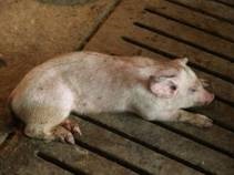
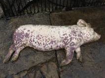
PCVD is generally seen in pigs from 6 to 14 weeks of age and was previously considered unlikely in pigs over 16 weeks of age although recent reports have described disease shortly after weaning and in heavy pigs. PCVD has been associated with Porcine Dermonephrotic Syndrome (PDNS) and reproductive failure and infection is presented clinically by mortality, ill- thrift (wasting), paleness, dyspnoea (difficulty breathing), intermittent diarrhoea and visibly enlarged lymph nodes.
PCVD is recognised as a multi-factorial disease and co-infections are considered one of the triggers in the disease onset. Subclinically infected pigs tend to be more susceptible to co-infections due to their compromised immune system.
Studies show that co-infections with Porcine reproductive and respiratory syndrome virus (PRRSV), swine influenza virus (SIV), porcine parvovirus (PPV), Mycoplasma hyopneumoniae, Actinobacillus pleuropneumoniae, Haemophilus parasuis and Streptococcus suis are commonly involved in PCVD.
Contributing factors
- Continuous population of houses
- High stocking rates
- Mixing and stress
- Low levels of biosecurity
- Poor hygiene (infected faeces not removed)
Clinical Signs of PCVD
The damage caused by PCVD depends on the amount of virus in growing pigs. High viraemia leads to clinical PCVD and potentially high mortality rates of up to 70 to 80% in affected animals, while low viraemia leads to less obvious subclinical disease, reduced average daily gain (ADG) and increased susceptibility to other pathogens.
While most herds would test positive for PCVD, the incidence of pigs infected with the virus that develop clinical signs of PCVD can be as low as 4%.
PCVD is characterised by
- Wasting/weight loss
- Paleness
- Intermittent diarrhoea
- Dyspnoea
- Enlarged inguinal lymph nodes
- Occasional jaundice
The weight loss is caused by nutrients intended for growth being redirected to the immune system and maintenance. There is a high mortality rate in affected piglets, most often within 48 to 72 hours, after onset of clinical signs. Some pigs survive a few weeks though recovery is rare if signs are severe. As a feature of PCVD immunosuppression pigs may also show signs of secondary bacterial or superimposed viral or fungal infections such as PRRS, E. coli enteritis, Salmonellosis and Haemophilis Parasuis (Glässers Disease).
Diagnosis
- Clinical signs
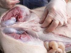
- Post mortem is an effective way of diagnosing PCVD
- Serology- ELISA based serology is only useful when paired sera, spaced 3 weeks apart, are tested showing seroconversion.
Treatment
As PCV2 is a virus, treatment with antibiotics is not considered an aid in the prevention or control of PCVD. Although some studies suggest that when bacterial infections are occurring simultaneously with PCV2 infection, marginal benefits may be gained from antibiotic treatment.
Control
Good management practices can have a significant effect on the control and spread of PCVD. The following approaches can assist in the control of disease:
- Strict bio-security and hygiene measures
- Strict adherence to all-in, all-out pig flow
- Removal and treatment of sick pigs from the group to a sick pen
- Minimal levels of cross fostering
- Avoid excessive stocking rates
Immunisation by vaccination of piglets from weaning is the most cost-effective method to control PCVD along with good management practices. The most popular vaccination regime is a one-dose regime at weaning. The chosen vaccination regime should provide protection against PCVD to the end of the fattening period which is usually at least 22 weeks.