Equine Respiratory Disease
A page about equine respiratory disease describing cause, clinical signs, diagnosis, and control.
Introduction
respiratory diseases are a serious problem in horses and depending on the causative agent, can lead to high morbidity and mortality rates. Furthermore, respiratory disease in horses can negatively impact the athletic potential of the horse. This effect is due to interruption of training and also the development of chronic debilitating disorders, for instance chronic pulmonary disease in untreated cases.
Respiratory diseases of horses are typically divided into conditions affecting the upper (i.e. the nose, throat and trachea) and the lower (i.e. the lungs) respiratory tract. The term inflammatory airway disease is a commonly used term to describe a type of respiratory condition where there is inflammation of the lower airways, with increased mucus and neutrophils. It is usually associated with exercise intolerance and poor performance in athletic animals. Some cases follow a viral infection. The initiating infection may even have been subclinical but allowed opportunistic infection to develop or caused airway hypersensitivity. Unfortunately due to the nature of the equine lifestyle many types of respiratory disease are common.
Aetiology
Various viral and bacterial agents can cause respiratory disease in horses and mixed infections are common. As is the case in other species, equine respiratory conditions often start with a viral infection, e.g. Equine Influenza, which may then be complicated by a secondary bacterial agent.
Epidemiology
Contributing factors to Equine Respiratory Disease:
1. Viruses- Several viruses are capable of initiating respiratory disease in horses: The most common of these viral pathogens in this region are;
Equine Influenza Virus- The causative agent of the respiratory disease in horses commonly known as “equine flu” (see “equine influenza” section).
Equine Herpes Virus- The subtypes of this virus most frequently associated with respiratory disease are subtypes 1 (EHV1) and 4 (EHV4). Equine herpesviruses are also associated with abortion, neonatal disease and neurological disease (see “equine herpesvirus” section).
2. Bacterial Agents- The most common organisms associated with pneumonia in horses are opportunistic bacteria originating from the resident flora of the upper respiratory tract. Secondary bacterial disease may result in mucosal bacterial infections (rhinitis and tracheitis) or may produce more serious invasive disease such as pneumonia and pleuropneumonia. Streptococcus equi zooepidemicus is the most common opportunistic pathogen of the equine lung, although Actinobacillus equuli, Bordetella bronchiseptica, Escherichia coli, Pasteurella spp, and Pseudomonas aeruginosa are frequently isolated.
Streptococcus equi var equi, the causative agent of strangles (see “strangles” section), is a primary bacterial pathogen of the upper respiratory tract and is capable of mucosal invasion without predisposing factors. Rhodococcus equi is also a primary pathogen of the lower respiratory tract of foals and produces pulmonary consolidation and abscessation. R. equi pneumonia is occasionally diagnosed in adult horses with a compromised immune system.
3. Environmental and Management- Exposure to organic dusts, particularly in older horses with a genetic predisposition to allergic airway disease, can lead to respiratory disease. Following exposure to allergens the small airways become obstructed by bronchoconstriction and excessive mucus production. This disease is known as Recurrent Airway Obstruction (RAO), formerly known as Chronic Obstructive Pulmonary Disease (COPD) or “heaves”. A recent UK study indicated that up to 1/3 of pony club horses and ponies may be affected by RAO.
Inflammatory airway disease, a common cause of exercise intolerance in young horses, is characterized by excessive mucus in the trachea and hyper reactivity of the airways. The initiating cause of the disease is unknown, but viral infections, allergy, and adverse environmental factors may facilitate the disease.
Exercise-induced pulmonary haemorrhage (EIPH) is a relatively common condition of sport horses and thoroughbred race horses that partake in strenuous exercise. The clinical findings reported with this condition are detection of haemorrhage during endoscopic examination or bronchoalveolar lavage and bleeding from one or both nostrils. It is widely believed that strenuous exercise causes the blood vasculature of the respiratory tract to change leading to haemorrhage in affected horses.
4. Parasitic- Lungworm, Dictyocaulus arnfeldi, is an important parasite of the lower respiratory tract of equidae. Clinical signs vary from asymptomatic infection to severe life threatening respiratory disease depending on the species infected, the number of lungworm larvae ingested, age and the immune status of the animal.
5. Neoplasia- Tumours of the respiratory tract in horses are rare but tumours may arise from any of the following structures in the respiratory tract: nasal and paranasal sinuses, nasopharynx, larynx, guttural pouches, lungs, pleura or thymus. Clinical signs vary according to the location of the tumour. For instance upper respiratory tract tumours often give rise to dyspnoea, nasal discharge and epistaxis whereas thoracic tumours may lead to poor oxygenation of the blood and cyanosis due to loss of lung capacity.
6. Miscellaneous-
Recurrent laryngeal hemiplegia occurs due to paresis or paralysis of the arytenoid cartilage and vocal folds. Affected horses are exercise intolerant and are often called ‘roarers’ due to the inspiratory respiratory noise they make during exercise.
Epiglottic entrapment is another cause of respiratory noise and exercise intolerance in the horse. Affected horses may also have a persistent cough and nasal discharge.
Dorsal displacement of the soft palate (DDSP) is another cause of respiratory noise and exercise intolerance in the horse. DDSP occurs when the caudal margin of the soft palate moves dorsal to the epiglottis which leads to a functional obstruction of the airway.
Fourth Branchial Arch Defect in horses is another performance limiting condition of the horse. The condition occurs due to the failure of extrinsic laryngeal structures to develop normally. Clinical signs of the condition include coughing, respiratory noise and difficulty in swallowing.
Nasal polyps can occur in horses of all ages and breed and can occur as singular or multiple pedunculated masses. Clinical signs in affected horses include inspiratory noise, nasal discharge and occasionally nasal haemorrhage. Occasionally the polyps may protrude from the affected nostril.
Disease of the paranasal sinuses typically results in nasal discharge, facial swelling and inspiratory noise. Causes of disease in the paranasal sinuses include sinusitis secondary to URT infections, ethmoid haematomas and sinus cysts.
Disease of the guttural pouches can result in a number of clinical signs including nasal discharge, nasal haemorrhage, pain in the parotid region, breathing difficulty, swallowing difficulty and even neurological disease. The most common causes of guttural pouch disease in the horse include empyema, mycosis and guttural pouch tympany.
Clinical signs
Clinical signs associated with respiratory disease vary according to the causative agent involved. However, respiratory disease associated with various viral agents have many similar clinical manifestations including pyrexia, serous nasal discharge, submandibular lymphadenopathy, anorexia, and cough. In addition to respiratory disease, equine herpesvirus type 1 (EHV-1) can cause abortion and neurological disease.
Clinical evidence of a bacterial infection includes mucopurulent nasal discharge, depression, persistent fever, abnormal lung sounds, hyperfibrinogenaemia and leucocytosis. Secondary bacterial disease may result in mucosal bacterial infections (rhinitis and tracheitis) or may produce more serious invasive disease such as pneumonia and pleuropneumonia.
Recurrent Airway Obstruction results in excessive mucus production and bronchoconstriction. The severity of clinical signs ranges from a mild cough to exercise intolerance and even severe dyspnoea at rest.
Diagnosis
A variety of diagnostic techniques in conjunction with the interpretation of clinical signs are used in the investigation of respiratory disease.
Swabbing of the nasopharynx, guttural pouches and lymph nodes can be a useful tool for virus isolation, bacterial culture and PCR.
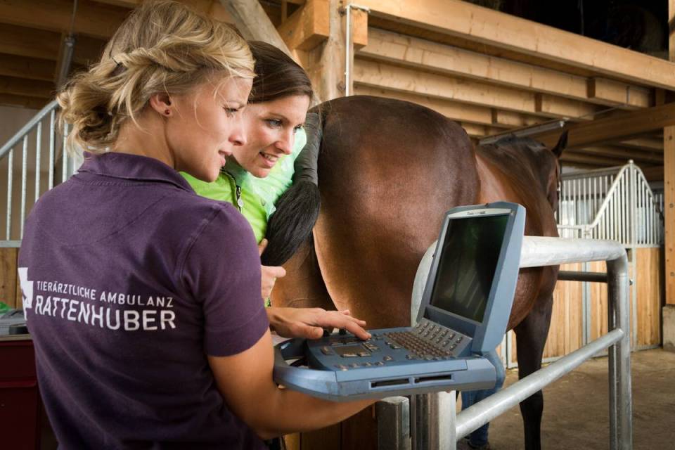
Endoscopic examination, commonly performed in racing thoroughbreds, allows visualization of the upper respiratory tract, guttural pouches, trachea, and main stem bronchi. Indications for endoscopic examination include upper airway noise, inspiratory difficulty, poor exercise performance, and unilateral or bilateral nasal discharge. Using the endoscope, bronchoalveolar lavage may be performed to collect a sample of fluid from the lower airways.
Transtracheal wash is a less commonly used technique to collect airway secretions for further analysis.
Radiographs of the skull may also be used to investigate sinus, guttural pouch and dental problems which may be associated with respiratory problems in horses.
Thoracic radiography and ultrasonography are valuable for assessing lower respiratory tract disease. Pulmonary consolidation (pneumonia), peribronchial disease, pulmonary abscessation, interstitial disease, and mediastinal masses (neoplasia, abscess, and granuloma) are most easily identified via thoracic radiography.
Thoracic ultrasonography is the most appropriate technique to evaluate fluid in the pleural space, peripheral pulmonary consolidation, and peripheral pulmonary abscessation. Ultrasonography can identify the volume, location, and character of pleural fluid or air within the pleural space (pneumothorax). Additionally, it can identify fibrin tags, gas echoes (anaerobic infection), masses, and fluid pockets. Ultrasound guided pleurocentesis is useful to obtain a fluid samples in the case of abnormal fluid build-up in the pleural space. This fluid can then be further analysed to determine the fluid type and culture may be performed on this fluid if bacterial infection is suspected.
Lung biopsy and fine needle aspiration are invasive procedures and performed only after other diagnostic procedures have been exhausted. Pulmonary neoplasia, pulmonary fibrosis, and interstitial diseases may require lung biopsy to obtain a diagnosis.
Treatment
Every possible effort should be undertaken to shorten the duration of the disease period as prolonged episodes of respiratory disease impact negatively on the horse’s welfare and if left untreated can lead to chronic disease sequelae.
As many cases are caused or complicated by bacterial infections, antibiotic therapy may be required. If possible the choice of the antibiotic should be based on culture and sensitivity of the bacteria present. If this is not possible then a broad-spectrum antibiotic that is effective against the likely pathogens involved may be chosen.
Antiparasitic drugs are indicated in the case of lungworm infection. Panacur Equine wormers are indicated for this condition.
Structural abnormalities, for example laryngeal hemiplegia and epiglottic entrapment, may require surgical intervention to repair.
Regardless of the type of respiratory disease, environmental factors and supportive care are important to aid recovery. A dust and ammonia-free (dry, urine free bedding) stable environment prevents further damage to the protective mechanisms of the respiratory system.
Highly palatable feeds are indicated to prevent weight loss during the treatment and recovery period. Adequate hydration will decrease the viscosity of respiratory secretions making the secretions easier to clear from the airways.
Rest is very important and affected horses should be kept in a dust-free environment, ideally by turning them out to pasture with no access to hay or straw. If this is not possible, then stabled horses should be bedded on shavings or shredded paper, and fed alternatives to hay, such as silage, or if hay is fed, it should be soaked prior to feeding.
In addition to antibiotics, the following drugs are used to provide symptomatic relief and hasten the resolution of infection:
• Bronchodilators,
• Mucolytics and expectorants
• Corticosteroids may be required in some cases of chronic lower airway inflammation. The use of corticosteroids should be avoided in active infections due to their immunosuppressive effect and long term steroid use can lead to laminitis.
• Non-steroidal anti-inflammatory drugs may be helpful in horses with pyrexia and inflammatory conditions of the respiratory tract.
Control
Vaccination is a crucial component in the control of respiratory disease in horses. Vaccines are used to immunise horses against Strep equi (“strangles”) and many of the viral infections associated with respiratory disease in the horse. Vaccination does not always prevent respiratory infections in horses, but duration and severity is usually lessened in horses that have been vaccinated regularly. Your veterinary practitioner can advise on an appropriate vaccination programme for your horse based on the probability of exposure and potential disease.
A variety of respiratory vaccines are available on the market either as single component vaccines like monovalent Influenza and Strep equi vaccines or as combined vaccines, for instance, combination influenza and tetanus vaccines. MSD Animal Health markets a variety of respiratory vaccine types. It is advisable to consult the SPC of the relevant product or your veterinary practitioner for advice on correct vaccination timing and regimes. For further information on equine respiratory disease and vaccination please click here.
The application of good hygiene, ventilation and management techniques is also vital to prevent respiratory disease in horses. Affected horses should be isolated until the causative agent is identified and treated appropriately. Maintain a closed yard until the infection is controlled. Ideally no horses should enter or exit the farm until outbreak is over to avoid the spreading of disease. Disinfect areas and equipment that have been used by infected and suspect horses, including stalls and feeders and remove as much organic waste materials as possible.
To prevent the introduction of disease to a naïve herd, newly acquired horses should be isolated for 4 weeks before introduction. In areas of risk, all newly acquired horses should have a complete and up to date vaccination history before introduction to the herd. Occasionally horses are swabbed for pathogens like Strep equi before introduction to the herd. These results should be interpreted with caution as some horses can give rise to false negative results.
Good hygiene and biosecurity in the yard are vital to minimise infections. Avoid stressors like overcrowding. Maintain disinfection of equipment and stables between horses. If travelling to events minimise mixing of horses from different yards and avoid sharing equipment with other horses.